New examination of fish considered a ‘living fossil’ changes our understanding of vertebrate skull evolution
Here is something that will cause creationists to jump for joy – until they read beyond the headline (if they ever do). Scientists have announced that they were wrong about the evolution of the vertebrate skull, including that of mammals.
However, beneath that headline lie some disappointing facts for creationists:
- The error was uncovered by re-examining the 400-million-year-old skull of a coelacanth.
- The mistake concerns details of how the vertebrate skull evolved – not whether it evolved.
- The paper directly contradicts the common creationist claim that scientists are only permitted to publish research that conforms to the scientific consensus. This study openly challenges the prevailing view.
- The discovery enhances our understanding of how the vertebrate skull evolved from that of ancestral lobe-finned fishes – precisely the kind of evidence creationists would rather didn’t exist.
Still, creationists can enjoy the headline and may even use it to 'prove' to their audience that science is unreliable because scientists sometimes make mistakes. Of course, they’ll likely ignore the fact that the fossil in question is 400 million years old, and gloss over the reality that – unlike religious dogma – science is a process of continuous refinement. Science allows for doubt, re-examination, and re-evaluation. When the evidence changes, scientists change their minds. In contrast, religious dogma is fixed and unchanging, usually despite the evidence, not because of it, hence the widening gap between what creationists are required to believe and what science reveals.
Upon re-examining the cranial musculature of the African coelacanth (Latimeria chalumnae), the researchers found that only 13% of the previously identified evolutionary muscle innovations in major vertebrate lineages were accurate. They also identified nine new evolutionary transformations related to innovations in feeding and respiration.
The researchers, Professor Aléssio Datovo from the University of São Paulo (USP) in Brazil and the late David Johnson from the Smithsonian Institution in the United States, who sadly died when the paper was in review, have just published their findings in Science Advances.
What can you tell me about coelacanths and their place in the evolution of vertebrates? Coelacanths are an extraordinary group of lobe-finned fish (Sarcopterygii) with an important place in vertebrate evolution. Often referred to as "living fossils," they provide crucial insights into the transition from aquatic to terrestrial vertebrates.They also discuss the significance of their findings in a press release by Fundação de Amparo à Pesquisa do Estado de São Paulo (FAPESP) by André Julião.
What Are Coelacanths?
Coelacanths belong to the order Coelacanthiformes, a lineage that was thought extinct until the first living specimen was discovered in 1938 off the coast of South Africa. This was Latimeria chalumnae; a second extant species, Latimeria menadoensis, was discovered in 1997 in Indonesia.
- They first appeared in the Devonian period, over 400 million years ago.
- Once a diverse and widespread group, only two known species survive today.
- Their closest relatives are lungfish and tetrapods (four-limbed vertebrates), not modern ray-finned fish.
Lobe-Finned vs Ray-Finned Fish
Coelacanths are lobe-finned fish, meaning their paired fins (pectoral and pelvic) are fleshy and limb-like, supported by a robust, bony internal skeleton.
- This is in contrast to ray-finned fish (Actinopterygii), whose fins are supported by long, thin bony rays.
- The structure of lobe fins is considered homologous to the limbs of terrestrial vertebrates, including humans.
Evolutionary Significance
Coelacanths are important in evolutionary biology because they:
- Represent a sister group to the lineage that gave rise to tetrapods – the first vertebrates to walk on land.
- Help scientists reconstruct the features of early vertebrates, especially around the skull, braincase, limb morphology, and internal anatomy.
- Possess several ancestral traits (plesiomorphies), including a notochord, intracranial joint, and a vestigial lung.
Key Contributions to Understanding Vertebrate Evolution:
- Fins to limbs: Their fin bones resemble those of early tetrapods.
- Cranial musculature and skull evolution: Recent work (like the paper you’re writing about) shows that coelacanths have retained primitive muscular structures that help clarify how cranial innovations arose in tetrapods.
- Vestigial lung: Provides evidence of a shared ancestry with lungfish and terrestrial vertebrates that had to breathe air.
Recent Research Implications The subject of this blog post highlights how re-examining the musculature of coelacanths has upended assumptions about vertebrate cranial evolution. This underscores their continued relevance in:Testing evolutionary hypotheses, Clarifying transitional morphologies, and Exposing limitations in past anatomical interpretations.
Ecology and ConservationCoelacanths live in deep-sea environments (100–500 metres), often in volcanic slopes or caves. They are nocturnal predators. Both known species are critically endangered, threatened by bycatch and habitat disturbance.
Summary: Coelacanths' Role in Vertebrate Evolution
- Lobe-finned anatomy: Illuminates the fin-to-limb transition
- Cranial structure: Sheds light on skull evolution in vertebrates
- Phylogenetic position: A close cousin to tetrapods, bridging aquatic and terrestrial life
- Retention of primitive traits: Offers a window into early vertebrate morphology
Here is a simplified cladogram showing the evolutionary relationship between coelacanths, lungfish, tetrapods, and ray-finned fishes:
- Coelacanths, lungfish, and tetrapods belong to the lobe-finned fishes (Sarcopterygii).
- Ray-finned fishes (Actinopterygians) are a separate lineage within vertebrates.
- Tetrapods (which include amphibians, reptiles, birds, and mammals) evolved from within the lobe-finned lineage, closely related to lungfish.

New examination of fish considered a ‘living fossil’ changes our understanding of vertebrate skull evolution
Researchers reanalyzed the skull musculature of coelacanths, a group of fish that has existed for 400 million years, and concluded that many structures had been incorrectly described. The study was published in Science Advances by researchers from the University of São Paulo and the Smithsonian Institution.
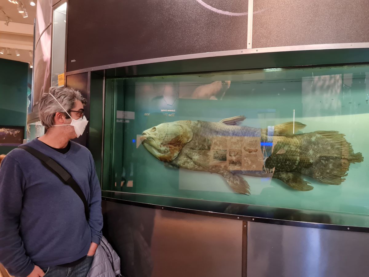
The coelacanth is known as a “living fossil” because its anatomy has changed little in the last 65 million years. Despite being one of the most studied fish in history, it continues to reveal new information that could transform our understanding of vertebrate evolution. This is revealed in a study published in the journal Science Advances by researchers from the University of São Paulo (USP) in Brazil and the Smithsonian Institution in the United States.
Upon re-examining the cranial musculature of the African coelacanth (Latimeria chalumnae), the authors discovered that only 13% of the previously identified evolutionary muscle novelties for the largest vertebrate lineages were accurate. The study also identified nine new evolutionary transformations related to innovations in feeding and respiration in these groups.
Ultimately, it’s even more similar to cartilaginous fish [sharks, rays, and chimaeras] and tetrapods [birds, mammals, amphibians, and reptiles] than previously thought. And even more distinct from ray-finned fish, which make up about half of living vertebrates.
Professor Aléssio Datovo, lead author
Museum of Zoology
University of São Paulo
São Paulo, SP, Brazil
Among the evolutionary novelties erroneously identified as present in coelacanths are muscles responsible for actively expanding the buccopharyngeal cavity, which extends from the mouth to the pharynx. This set of muscles is directly related to food capture and respiration. However, the study showed that these supposed muscles in coelacanths were actually ligaments, which are structures incapable of contraction.
Ray-finned fish (actinopterygii) and lobe-finned fish (sarcopterygii) diverged from a common ancestor approximately 420 million years ago. The sarcopterygii include fish such as coelacanths and lungfish, as well as all other tetrapods, because they evolved from an aquatic ancestor. These include mammals, birds, reptiles, and amphibians.
In ray-finned fish, such as aquarium carp, it is easy to see how the mouth moves to suck in food. This ability gave actinopterygii a significant evolutionary advantage; today, they comprise about half of all living vertebrates.
This is a fundamental difference from other fish, such as coelacanths and sharks, which primarily feed by biting their prey.
In previous studies, it was assumed that this set of muscles that would give greater suction capacity was also present in coelacanths and, therefore, would have evolved in the common ancestor of bony vertebrates, which we now show isn’t true. This only appeared at least 30 million years later, in the common ancestor of living ray-finned fish.
Professor Aléssio Datovo.
Behind the scenes
Coelacanths are extremely rare fish that live about 300 meters below the surface of the water and spend their days in underwater caves.
One reason they have changed so little since the extinction of the dinosaurs is that they have few predators and live in a relatively protected environment. This has resulted in slow changes to their genome, as shown by a 2013 study published in the journal Nature.
Coelacanths were first known only from fossils from about 400 million years ago. It was not until 1938 that a living animal was discovered, much to the astonishment of scientists. In 1999, another species (Latimeria chalumnae) was discovered in Asian waters.
Due to the rarity of specimens in museums, researchers from USP and the Smithsonian Institution’s National Museum of Natural History had to persevere to find an institution willing to lend animals for dissection.
The Field Museum in Chicago and the Virginia Institute of Marine Science, both in the United States, finally agreed to lend one specimen each. According to Datovo, G. David Johnson, co-author of the article, deserves credit for obtaining the loan.
Johnson, born in 1945, was “probably the greatest fish anatomist of his time,” according to Datovo. He died in November 2024 after a domestic accident while the study was under review.
Contribution
Contrary to what it may seem, dissecting a specimen does not mean destroying it as long as it’s done properly.
Professor Aléssio Datovo.
The researcher, who has been conducting this type of study for over 20 years, spent six months separating all the muscles and skull bones of the coelacanth. These structures are now preserved and can be studied individually by other scientists, eliminating the need to dissect a new animal.
Seeing each muscle and nerve firsthand allowed the authors to identify what was actually in the coelacanth’s head with certainty, point out previously undescribed structures, and correct errors that had been repeated in the scientific literature for over 70 years.
There were many contradictions in the literature. When we finally got to examine the specimens, we detected more errors than we’d imagined. For example, 11 structures described as muscles were actually ligaments or other types of connective tissue. This has a drastic consequence for the functioning of the mouth and breathing, because muscles perform movement, while ligaments only transmit it.
Professor Aléssio Datovo.
Due to the position of coelacanths in the vertebrate tree of life, the discovery impacts our understanding of cranial evolution in all other large vertebrate groups.
With this information, the researcher used three-dimensional microtomography images of the skulls of other groups of fish, both extinct and living. These images are made available by other researchers who study fish anatomy when they perform 3D scans.
From images of the skull bones of other fish from completely extinct lineages, Datovo and Johnson were able to infer where the muscles found in coelacanths would fit, elucidating the evolution of these muscles in the first jawed vertebrates. In future work, Datovo intends to analyze similarities with the muscles of tetrapods, such as amphibians and reptiles.
Publication:
AbstractThis discovery is particularly bad news for creationists, not because it undermines evolutionary biology — it doesn't; it confirms it — but because it directly contradicts several of their most cherished talking points. Far from being an embarrassment for science, this is an example of science working exactly as it should: through re-examination, correction, and refinement. The fact that scientists identified and corrected a long-standing misconception about vertebrate skull evolution, based on fresh analysis of a 400-million-year-old fossil, is a testament to the self-correcting nature of the scientific method.
Coelacanths are rare fishes that occupy a key evolutionary position in the vertebrate tree of life. Despite being exhaustively studied, we found that a substantial part of the knowledge on their cranial musculature was mistaken. Eleven previously reported coelacanth “muscles” are nonexistent, while three previously unknown muscle subdivisions and connections are found. These findings markedly affect our understanding of the deep-time cranial evolution of jawed vertebrates (gnathostomes). Only 13% of the previously identified myological evolutionary novelties for the major gnathostome lineages proved to be accurate, but several new ones are proposed. We show that low, moderate, and high levels of cranial muscle innovation characterized the emergence of lobe-finned (sarcopterygian), cartilaginous (chondrichthyan), and ray-finned (actinopterygian) fishes, respectively. The novelties in the latter group resulted in the evolution of a second active mechanism for the expansion of the oropharyngeal cavity, which was probably crucial for the predominance of suction feeding versus bite feeding in extant actinopterygians.
INTRODUCTION
Gnathostomes comprise over 99% of extant vertebrate diversity. Enormous progress has been made in understanding the major skeletal evolutionary transformations within this group. However, the deep-time evolution of their muscular system remains elusive. Part of this problem stems from poor knowledge of muscle homologies across large groups (1, 2). Our study markedly illustrates a second problem: the lack of reliable information about musculature in the literature. Despite being one of the most iconic living vertebrates, we found a plethora of errors in the identification of cranial muscles in the African coelacanth (Latimeria chalumnae), some of which have been replicated for nearly 70 years. Given the key position of coelacanths in the vertebrate tree of life, correcting these errors has profound implications for understanding the early gnathostome evolution.
Fig. 1. Cranial musculoskeletal system of African coelacanth, L. chalumnae, with associated motor branches of cranial nerves.
FMNH 76057 (CCC 59), 1070-mm total length (TL). Left lateral view with ocular muscles, facial bones, and mandibulo-hyoid ligaments removed; outlines of opercle, spiracular ossicle, and dorsal portions of sphenoidalis and rictalis represented by dashed lines. Red diamonds indicate structures unreported or erroneously reported in past studies (see the Results). CHD, constrictor hyoideus dorsalis; CHV, constrictor hyoideus ventralis.Fig. 2. Connection diagrams of mandibular muscles in major gnathostome lineages.
Expansor and compressor muscles in blue and red, respectively. A, adductor palatoquadrati; B, buccalis segment of AM; C, constrictor mandibularis dorsalis; D, dilatator operculi; Eth, ethmoid region; F, facialis segment of AM; Hym, hyomandibular; L, levator arcus palatini; LwJ, lower jaw; Ope, opercle (=submarginal plate in placoderms); N, sphenoidalis section of AM; O, orbitalis section of AM, P, levator palatoquadrati; Paq, palatoquadrate; PoF, postorbital fossa; PoP, postorbital process; Pro, preopercle; S, spiracularis; Spr, spiracle and/or spiracular ossicle or cartilage.Fig. 3. Attachment sites and reconstructions of mandibular muscles in sarcopterygians.
Actinistian L. chalumnae, (A) left lateral view of cranium [redrawn from figure 1 in (60)]. Porolepiform Durialepis edentatus, (B) medial and (C) lateral views of left palatoquadrate and dermopalatines 61) and (D) left lateral view of schematic reconstructions of mandibular muscles [skeleton based on figure 9 in (12); https://creativecommons.org/licenses/by/4.0/deed.en; endoskeletal elements in gray and separation between superficial bones in dotted lines]. Not to scale.Fig. 4. Attachment sites and reconstructions of mandibular muscles in chondrichthyans. Hexanchiform Chlamydoselachus anguineus, left lateral views of (A) neurocranium, mandibular, and hyoid arches [redrawn from figure 7 in (45); https://creativecommons.org/licenses/by/4.0/deed.en] and (B) schematic reconstructions of mandibular muscles. Symmoriiform Ferromirum oukherbouchi, (C) left lateral view of the neurocranium, mandibular, and hyoid arches [redrawn from figure 4 in (39); https://creativecommons.org/licenses/by/4.0/]. Cladoselachiform Maghriboselache mohamezanei, (D) left lateral view of schematic reconstructions of mandibular muscles [skeleton based on figure 5 in (38); https://creativecommons.org/licenses/by/4.0/]. Not to scale.Fig. 5. Attachment sites and reconstructions of mandibular muscles in acanthodians.
Acanthodiform Acanthodes bronni, left lateral views of (A) part of braincase and (B) schematic reconstructions of mandibular muscles [skeleton redrawn from figure 4.7 in (34); https://creativecommons.org/licenses/by/4.0/deed.en]. Acanthodiform Acanthodes confuses, (C) lateral view of left palatoquadrate and lower jaw and (D) medial view of left palatoquadrate [redrawn from figure 6 in (21); https://creativecommons.org/licenses/by-nc/4.0/legalcode.en]. Not to scale.Fig. 6. Attachment sites and reconstructions of mandibular muscles in placoderms.
Buchanosteid arthrodire ANU V244, (A) ventral view of braincase, (B) medial and (C) dorsal views of right palatoquadrate and associated dermal plates, and (D) lateral view of left Meckel’s cartilage [redrawn from figures 3, 4, and 6 in (23); https://creativecommons.org/licenses/by/4.0/]. Arthrodiran Coccosteus cuspidatus, (E) left lateral view of schematic reconstructions of mandibular muscles [endoskeletal elements in gray and separation between superficial bones in dotted lines; skeleton based on figure 7 in (62)]. Not to scale.Fig. 7. Attachment sites and reconstructions of mandibular muscles in actinopterygians.
Polypteriform Polypterus bichir, left lateral views of (A) braincase and (B) suspensorium, and (C) medial view of right opercle [redrawn from figures 7, 29, and 33 in (63)]. Stem actinopterygian Raynerius splendens, (D) lateral and (E) dorsal views of left palatoquadrate and associated preopercle fragments (64). Stem actinopterygian Mimipiscis toombsi, (F) left lateral view of schematic reconstructions of mandibular muscles [endoskeletal elements in gray and separation between superficial bones in dotted lines; skeleton based on figure 101 in (32); https://creativecommons.org/licenses/by-nc-sa/4.0/]. Stem actinopterygian Australosomus kochi, (G) cross section at middle of maxillary-palatoquadrate chamber [redrawn from figure 31 in (28)]. Not to scale.Fig. 8. Evolutionary transformations (maximum parsimony) of cranial muscles mapped over simplified phylogenetic tree of major gnathostome lineages.
Topology based on [figure 6 in (22) and figure 11 in (27); https://creativecommons.org/licenses/by/4.0/deed.en]. Miniatures filled with gray indicate extant taxa. Synapomorphies: 1, levator palatoquadrati subdivided into levator arcus palatini and dilatator operculi; 2, CMD inserting on lateral face of palatoquadrate (10); 3, CMD inserting on hyomandibular (10); 4, presence of adductor palatoquadrati; 5, facialis originating from preopercle; 6, facialis originating from hyomandibular; 7, orbitalis originating from ethmoid region; 8, extrapalatoquadrate ridge surpassing postorbital process and coopting origin of sphenoidalis; 9, sphenoidalis originating from ethmoid region; 10, buccalis originating from neurocranium; 11, absence of CBS; 12, coracomandibularis originating from hypobranchial 3 (8, 9); more details about characters in the Results and the Supplementary Materials.
Aléssio Datovo, G. David Johnson
Coelacanths illuminate deep-time evolution of cranial musculature in jawed vertebrates.
Sci. Adv. 11, eadt1576 (2025). DOI:10.1126/sciadv.adt1576
Copyright: © 2025 The authors.
Published by the American Association for the Advancement of Science. Open access.
Reprinted under a Creative Commons Attribution 4.0 International license (CC BY 4.0)


Creationists often portray science as dogmatic, claiming that scientists are only allowed to publish findings that align with an evolutionary “agenda.” But this study, which challenges prevailing views and overturns widely accepted assumptions, completely dismantles that myth. It was published in a leading peer-reviewed journal precisely because it presented robust evidence that prompted a re-evaluation of existing ideas. Far from being suppressed, it was celebrated — as real science always is when it moves our understanding forward.
Yet, predictably, creationists will seize on the headline — “Scientists were wrong!” — and use it to peddle their favourite narrative: that science is unreliable because it sometimes makes mistakes. What they won't tell their audience is that this process of self-correction is science’s greatest strength, not a weakness. They will ignore the fact that the fossil in question is 400 million years old, and that the findings further reinforce — not contradict — the reality of evolution. Unlike religious dogma, which clings rigidly to unchanging doctrine regardless of evidence, science welcomes change when the facts demand it. And that, ultimately, is what makes creationism so intellectually bankrupt in comparison.
The also shows scientists, far from abandoning the Theory of Evolution in favour of creationism, as many creationists have been tricked into believing, but explaining how their discovery improves our understanding of the evolutionary process. The fossils are of course, transitional, just as the Theory of Evolution predicts.
Advertisement
What Makes You So Special? From The Big Bang To You

Ten Reasons To Lose Faith: And Why You Are Better Off Without It


All titles available in paperback, hardcover, ebook for Kindle and audio format.
Prices correct at time of publication. for current prices.

No comments:
Post a Comment
Obscene, threatening or obnoxious messages, preaching, abuse and spam will be removed, as will anything by known Internet trolls and stalkers, by known sock-puppet accounts and anything not connected with the post,
A claim made without evidence can be dismissed without evidence. Remember: your opinion is not an established fact unless corroborated.