
The problems for the creation cult continue to pile up, no-doubt behind the backs of creationists who habitually look the other way when science produces more evidence refuting their dogmas.
This time, it's yet more evidence of the origins of terrestrial tetrapods, including mammals, as air-breathing fish. The evidence is that the basic structures of the mammalian shoulder joint were already present in a fossil fish, and it closes another of creationism beloved gaps in which they force-fit their ever-shrinking little god.
The shoulder girdle – the configuration of bones and muscles that in humans support the movement of the arms – was a classic example of an evolutionary ‘novelty’. This is where a new anatomical feature appears without any obvious precursors; where there is no smoking gun of which feature clearly led to another.
The new study uses fossils, developmental biology, and comparative anatomy, to investigate the 'mystery' and in so doing suggests a new approach to investigating the evolution of major anatomical structures, like the shoulder girdle.
The fossil studied was of the late Devonian placoderm fish, Kolymaspis sibirica:
What information do you have on the archaic fish, Kolymaspis sibirica and its place in the evolution of terrestrial vertebrates? Kolymaspis sibirica is an extinct, ancient fish species that lived during the Late Devonian period, approximately 375 million years ago. It is considered a member of the group of fishes known as Placodermi, which are often referred to as "armored fishes" due to the presence of bony plates covering their bodies. Placoderms were a diverse group of fishes that existed from the Silurian to the end of the Devonian period, and they played a significant role in the evolutionary history of vertebrates.The study was led by Dr Martin Brazeau of Imperial College London and researchers from The Natural History Museum, London. Their results are published in Nature.
Kolymaspis sibirica, like other placoderms, possessed a heavily armored exoskeleton, which provided protection and support for its body. It had a flattened body shape and was adapted for a bottom-dwelling lifestyle. Some of the key features of Kolymaspis sibirica include its large, jawed mouth and the presence of paired pectoral and pelvic fins.
In terms of the evolution of terrestrial vertebrates, the placoderms, including Kolymaspis, are of interest because they represent a stage in vertebrate evolution when certain adaptations started to appear that would later become important for the transition from aquatic to terrestrial life. While placoderms themselves were fully aquatic, their descendants eventually gave rise to the earliest amphibians, which were the first vertebrates to successfully transition to land. Some of the key adaptations that can be traced back to placoderms like Kolymaspis and are important in the evolution of terrestrial vertebrates include:It's important to note that while placoderms like Kolymaspis sibirica played a role in the evolutionary history of terrestrial vertebrates, they did not directly give rise to amphibians or other tetrapods. The transition to land involved many more steps and adaptations over millions of years. Nonetheless, the study of placoderms and their characteristics helps scientists understand the gradual development of traits that facilitated life on land, eventually leading to the diverse group of terrestrial vertebrates we see today.
- Paired Fins: Placoderms, including Kolymaspis, had paired pectoral and pelvic fins, which are homologous to the limbs of tetrapods (four-limbed vertebrates). These fins provided greater maneuverability and could have been precursors to limbs.
- Bony Armor: Placoderms had bony plates covering their bodies. Over time, some descendants of placoderms evolved thicker, more robust bones in their fins, which eventually contributed to the development of limbs.
- Breathing Adaptations: As aquatic animals, placoderms had gills for underwater respiration. In the transition to terrestrial life, air-breathing structures, such as lungs, evolved from primitive gills, and this change was crucial for terrestrial vertebrates.
The research and its significance are described in an Imperial College press release by Hayley Dunning:
Foundations of fins
One theory of the shoulder’s origin is that it was part of how fins formed in pairs on either side of the fish body, the evolution of which allowed fish more swimming control and eventually spurred the move from water to land.
The ‘gill-arch’ hypothesis suggests that these fins evolved from the bony ‘loops’ that support the gills, which also formed the shoulder. However, it has been difficult to gather any evidence for this hypothesis, as the features are rarely preserved in fossils.
A different theory of how the fins formed, the ‘fin-fold’ hypothesis, suggests the precursors of the paired fins instead evolved out of a line of muscle on the flanks of the fish. This theory has gained a lot of supportive evidence in the 150 years since both were proposed, but it cannot explain how the associated shoulder girdle evolved.
Now, by reanalysing an ancient fossil fish skull from soon after the shoulder girdle emerged, alongside other lines of evidence, the team suggest the truth may lie in a modified version of the gill-arch hypothesis that reconciles it with the fin-fold hypothesis.
Inside the brain case
The fossil the team looked at is a placoderm, of the species Kolymaspis sibirica, which lived around 407 million years ago and was among earliest jaw-bearing fishes. The fossil has a well-preserved brain case – the hard inner parts of the skull that record imprints and other features of the brain.
Dr Brazeau realised that despite the poor or absent preservation of the gill arches in such fossils, evidence for them could be well preserved in the brain case: the cartilaginous or bony ‘box’ that surrounds the brain and supports the sensory structures like eyes and ears. The brain case showed a curious head-shoulder joint highlighted by the configuration of muscles and blood vessels.
By comparing this feature in the jawed fish fossil with the brain case features of their precursors, the jawless fish, he and the team discovered new ways the two could be compared. They found the unusual head-shoulder joint bears similarities with the gill arches in earlier fish, suggesting it was these that were retained and incorporated into the formation of the shoulder at an early stage.
While most jawless fish have 5-20 gill arches, jawed fish almost never have more than five. Combining this with the new brain-case evidence, the team suggest the sixth gill arch was incorporated into the shoulder, becoming a crucial boundary that separated the head from the body. Intriguingly, the blood supply to the fins of jawless fishes emerges between the sixth and seventh gill arches.
Moving bones
This finding also means it doesn’t have to be an either/or in terms of how the paired fins evolved. Dr Brazeau added: “Our study shows how there is merit to both theories without accepting one or the other wholesale. Instead, we can rationalise the areas that overlap.”The gill arches seem to have been involved in the early separation of the head and body via the shoulder. But we no longer have gill arches – though the shoulder was templated on them, they don’t need to still be around today. This is consistent with some earlier studies that showed muscles can remain highly stable, while the specific bones that support them gradually take over one from the other. Gill arches may have done their part and been replaced as the shoulder took on a new configuration, including supporting things like our necks.
Martin D. Brazeau, lead author
Department of Life Sciences
Imperial College London, Ascot, UK
And The Natural History Museum, London, UK.
Dr Zerina Johnson, Researcher at the Natural History Museum, added: “The team will next focus on specimens from the Natural History Museum’s fossil fish collection. This will include jawless fish that have fins but lack a distinct shoulder girdle.
“We are currently processing many gigabytes worth of data, and I can hardly wait to see what these important specimens from the collection will add to the story.”

A detailed description and more illustrations are provided in the team's open access paper in Nature:
AbstractTo recap: we have here a fossil fish, Kolymaspis sibirica, that lived 407 million years before 'Creation Week' which shows distinct evidence of transition from the gill-arches of a primitive fish to a shoulder girdle, as a precursor, firstly to walking on the sea bed and later to crawling out onto land and eventually becoming the bones of the shoulder girdle to which the front limbs are attached. This fills another one of those gaps in which creationists try to fit their shrinking little god. And once again, when science closes a gap, no gods were needed in the explanation and the Theory of Evolution, which predicts these transitional fossils, was proved correct again.
The origin of vertebrate paired appendages is one of the most investigated and debated examples of evolutionary novelty1,2,3,4,5,6,7. Paired appendages are widely considered as key innovations that enabled new opportunities for controlled swimming and gill ventilation and were prerequisites for the eventual transition from water to land. The past 150 years of debate8,9,10 has been shaped by two contentious theories4,5: the ventrolateral fin-fold hypothesis9,10 and the archipterygium hypothesis8. The latter proposes that fins and girdles evolved from an ancestral gill arch. Although studies in animal development have revived interest in this idea11,12,13, it is apparently unsupported by fossil evidence. Here we present palaeontological support for a pharyngeal basis for the vertebrate shoulder girdle. We use computed tomography scanning to reveal details of the braincase of Kolymaspis sibirica14, an Early Devonian placoderm fish from Siberia, that suggests a pharyngeal component of the shoulder. We combine these findings with refreshed comparative anatomy of placoderms and jawless outgroups to place the origin of the shoulder girdle on the sixth branchial arch. These findings provide a novel framework for understanding the origin of the pectoral girdle. Our evidence clarifies the location of the presumptive head–trunk interface in jawless fishes and explains the constraint on branchial arch number in gnathostomes15. The results revive a key aspect of the archipterygium hypothesis and help reconcile it with the ventrolateral fin-fold model.
Fig. 1: The braincase and skull roof of K. sibirica Bystrow 1956 specimen TsNIGR 7656 as a virtual three-dimensional rendering.
 a, Dorsal view. b, Ventral view. c, Interpretive illustration of ventral view. d, Left lateral view. e, Posterior view. a.ic, foramen for internal carotid artery; art.crs, articular facet on end of craniospinal process; art.fac, articular facets for branchial arches; crs.p, craniospinal process; cu.fo, cucullaris muscle fossa; eyst, eystalk attachment; fo.mag, foramen magnum; gle.fo, fossa for occipital glenoid facets; hyp.fo, hypophyseal fossa; lba, laterobasal angle; N.II, optic tract canal; na, naris; not.c, notochordal canal; o.dend, endolymphatic duct opening; o.pin, pineal opening; orb.l, left orbit; orb.r, right orbit; Prm, premedian plate; rhi.fi, rhinocapsular fissure. Dark beige material is dermal (exoskeletal) bone and light beige material is perichondral (endoskeletal) bone.
Fig. 2: Comparative anatomy of cranial processes and branchial arch attachments in stem gnathostomes.
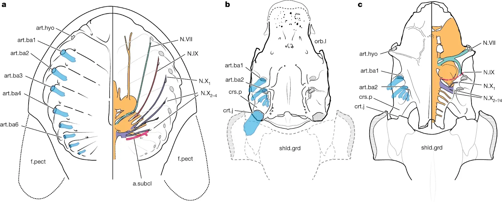 a, Osteostracan Nectaspis (composite based on ref. 47). b, Acanthothoracid placoderm Kolymaspis. c, Acanthothoracid placoderm Romundina (original based on data from ref. 48 and new data). Transparent blue structures represent reconstructed branchial arches. art.ba1–6, serially numbered branchial arch attachments (corresponds to art.fac in Fig. 1); art.hyo, hyoid arch articulation; a.subcl, canal for subclavian artery; crt.j, craniothoracic joint; f.pect, pectoral fin; N.VII, facial nerve canal; N.IX, glossopharyngeal canal; N.X1–4, vagus nerve canal branches (numbered 1–4); shld.grd, shoulder girdle. Not to scale.
Fig. 3: Summary phylogeny of early gnathostomes with reconstructions to show comparative anatomy of pharyngeal arches and shoulder linkages.
 Hypothetical intermediate is shown, for clarity of comparative anatomy; specific geometries may have varied substantially. Gill arch morphologies in osteostracan and placoderms are hypothetical and are shown to indicate location of articulations and constraints on overall pharynx architecture. Blue, branchial arches; orange, sixth branchial or thoracic arch; pink, pectoral fin attachment or scapulocoracoid. See Supplementary Information for complete phylogeny. Dashed lines indicate inferred pectoral girdle. Osteostracan is a composite based on ref. 47; Romundina is based on ref. 49 and new data; Eusthenopteron is a composite based on ref. 50.Brazeau, M.D., Castiello, M., El Fassi El Fehri, A. et al.
Fossil evidence for a pharyngeal origin of the vertebrate pectoral girdle. Nature (2023). https://doi.org/10.1038/s41586-023-06702-4
Copyright: © 2023 The authors.
Published by Springer Nature Ltd. Open access.
Reprinted under a Creative Commons Attribution 4.0 International license (CC BY 4.0)
And yet creationism staggers on and frauds continue to fleece suckers for a living by pandering to the cult's clamour for disinformation.

No comments :
Post a Comment
Obscene, threatening or obnoxious messages, preaching, abuse and spam will be removed, as will anything by known Internet trolls and stalkers, by known sock-puppet accounts and anything not connected with the post,
A claim made without evidence can be dismissed without evidence. Remember: your opinion is not an established fact unless corroborated.