

Illustration of Naegleriavirus based on electron microscopy. A section through a virus particle with the star-shaped stargate is shown.
Credit: Stefan Pommer / photopic.at (CC BY-NC-SA 4.0)
Judging by the findings of scientists led by Patrick Arthofer and Matthias Horn from the University of Vienna's Center for Microbiology and Environmental Systems Science (CeMESS), creationism's divine malevolence may have gone entirely round the twist and got carried away with its success with parasites.
Not only has it created parasites, apparently to increase the suffering in the world, but it's now creating parasites for those parasites! Kindly creationists who have realised blaming another creator such as 'Sin' for parasites is a blasphemy, might be tempted to credit their beloved pestilential malevolence with trying to combat the parasites like the single-celled organism, Naegleria fowleri, that are causing such misery by attacking them with another parasite. But that makes no sense in terms of an omniscient, omnipotent creator god who, if such a god existed, could simply destroy Naegleria fowleri and have done with it.
The only thing that makes sense, if you believe the childish notion of intelligent design by a magic invisible man is that it has become obsessed with creating parasites for the sake of creating parasites.
What the Vienna team have discovered is that the serious pathogen, Naegleria fowleri, is parasitised by a giant virus, named Naegleriavirus. Giant viruses are unusually large viruses with a complex genome.
Naegleria fowleri is an especially nasty little amoeba that I described in my illustrated book, The Malevolent Designer: Why Nature's God is not Good:
Naegleria fowleri is an amoeba which destroys the human brain, but it doesn't do it simply by eating it - that would be too simple for our malevolent designer. It does it by making us destroy our own brain, not just because our immune system is not fit for purpose but also because of the way our brain is fitted into our skull.
N. fowleri lives in warm, freshwater pools, normally living on a diet of local bacteria. However, should someone take a dip in one of these pools and gets water up their nose, some of the amoebas might penetrate the mucous membrane and find their way up the nerves to the brain, where they start eating brain cells, causing amoebic encephalitis.
Thankfully, this is a reasonably rare event because it is almost always fatal. Of 132 people in the USA known to have been infected since 1962, only three survived.
Infection is more common elsewhere: in Pakistan, some 20 people a year die from infection by N. fowleri. But the amoeba itself is almost certainly not what actually kills people.
According to a paper published in Acta Tropica by Abdul Mannan Baig from Aga Khan University in Karachi, Pakistan, the main culprit could be the host's immune system itself which does most of the damage (35).
The immune system's response is to flood the brain with immune cells. Not only do the enzymes released by the immune cells damage the brain cells themselves, but this response causes inflammation and swelling. Normally, swelling and inflammation around an infection site are exactly what's needed because this brings more blood and more lymphocytes to the area, helping to fight the infection and promote healing. In the brain, however, this response can be disastrous.
The problem is a piece of really bad ‘design’. The brain is contained in a bony case which can't expand and from which there is only one significant outlet through which pressure build-up can be dissipated - the foramen magnum, i.e., the hole through which the brain stem passes to form the spinal cord. Basically, when the brain swells, it is like trying to squeeze toothpaste out of the nozzle. The result is compression of the brain stem by a process the medical profession calls 'coning'. And this is where the second piece of bad design comes in.
All the deep centres needed to maintain basic life-support such as respiration, blood pressure and heart rate are located in the brainstem and get knocked out by coning because the pressure closes the supplying blood vessels.
Abdul Mannan Baig, of the Department of Biological and Biomedical Sciences at Karachi University, believes he has shown that brain cells actually survive longer without an immune response anyway, so he recommends a treatment for amoebic encephalitis which amounts to over-riding the 'Intelligent Design' of the body and suppressing the body's immune system before hitting the parasites with specific drugs. This should help reduce the risks from brain-swelling too.
Rosa Rubicondior - The Malevolent Designer: Why Nature's God is not Good, pp. 32-33
But, if you believe in the creationist superstition, this isn't so much about one of your divine malevolence's nasty little parasites but about a virus that it has designed to live on its parasite (are you still with me?)
That is the subject of an open access paper in Nature Communications and research which is described in a University of Vienna News release. The way the virus kills it's host it typical of the sneaky designs by creationism's designer - it suppresses the hosts immune response, using proteins coded for by genes it stole from its host species, in order to keep it alive and use its resources until it has successfully created enough new viruses, then it kills it and releases more viruses to kill more host cells:
New unusual giant virus discovered in wastewater treatment plant near Vienna
The single-celled organism Naegleria fowleri ranks among the deadliest human parasites. Researchers around Matthias Horn and Patrick Arthofer from the Center for Microbiology and Environmental Systems Science at the University of Vienna, in an international collaboration, have discovered viruses that infect this harmful microbe. Named Naegleriavirus, these belong to the giant viruses, a group known for their unusually large particles and complex genomes. The team details their findings in the prestigious journal, Nature Communications. Naegleri species are single-celled amoebae, found globally in water bodies. Notably, one species, Naegleria fowleri, thrives in warm waters above 30°C and causes primary amoebic meningoencephalitis (PAM), a rare but almost invariably fatal brain infection. A research team led by Patrick Arthofer and Matthias Horn from the University of Vienna's Center for Microbiology and Environmental Systems Science (CeMESS) has now isolated giant viruses that infect various Naegleria species. Giant viruses, scientifically termed Nucleocytoviricota, are a virus group identified just two decades ago, primarily infecting single-celled organisms. These viruses rival bacteria in size, boasting unique structures and genetic traits previously thought exclusive to cellular life. Their discovery sparked debates over the definition of viruses and the origins of life.This discovery and the characterization of Naegleriaviruses were made possible through international collaboration with researchers from the universities in Poitiers, the Canary Islands, and the US-based Joint Genome Institute.The newly discovered Naegleriaviruses were isolated from a waste water treatment plant in Klosterneuburg near Vienna and represent only the fourth isolate from a group called Klosneuviruses.
Patrick Arthofer, first author
Centre for Microbiology and Environmental Systems Science
Division of Microbial Ecology
University of Vienna, Vienna, Austria.
Naegleriaviruses are taken up mistakenly as a food source but destroy their amoeba hosts within just few hours. They exhibit a structure familiar in giant viruses, infecting host cells via a so-called stargate structure that facilitates DNA entry. Within hours, a structure known as a virus factory forms inside the amoeba cell, replicating viral genetic material outside the nucleus and assembling hundreds of new virus particles. To keep the host cell alive during this process, Naegleriaviruses likely use special proteins that suppress the cell's natural immune response, preventing premature cell death. Only after successful viral replication does cell destruction and virus release occur.
Viruses are employed in phage therapy to combat bacterial pathogens. "The newly identified Naegleriaviruses may not be suitable to treat Naegleria infections, given the challenging accessibility of the brain, where infections occur. However, this discovery opens the door to the possibility of preventative treatment of at-risk water bodies, such as during swimming pool water treatment, but this would first require further research. Regardless, the discovery of these viruses will enhance our understanding of both Naegleria biology and the viruses that infect them," says Matthias Horn.
AbstractIt is things like this that highlight better than anything the stark contrast between what we would expect from a nature designed by an omnibenevolent, omniscient super-intelligent designer and one we would expect from a mindless, utilitarian evolutionary process. The only sane way to understand parasites, and parasites on parasites is if they are a product of the latter, since no intelligence, malevolent or compassionate would design something so obviously and spectacularly stupid as this.
Giant viruses (Nucleocytoviricota) are significant lethality agents of various eukaryotic hosts. Although metagenomics indicates their ubiquitous distribution, available giant virus isolates are restricted to a very small number of protist and algal hosts. Here we report on the first viral isolate that replicates in the amoeboflagellate Naegleria. This genus comprises the notorious human pathogen Naegleria fowleri, the causative agent of the rare but fatal primary amoebic meningoencephalitis. We have elucidated the structure and infection cycle of this giant virus, Catovirus naegleriensis (a.k.a. Naegleriavirus, NiV), and show its unique adaptations to its Naegleria host using fluorescence in situ hybridization, electron microscopy, genomics, and proteomics. Naegleriavirus is only the fourth isolate of the highly diverse subfamily Klosneuvirinae, and like its relatives the NiV genome contains a large number of translation genes, but lacks transfer RNAs (tRNAs). NiV has acquired genes from its Naegleria host, which code for heat shock proteins and apoptosis inhibiting factors, presumably for host interactions. Notably, NiV infection was lethal to all Naegleria species tested, including the human pathogen N. fowleri. This study expands our experimental framework for investigating giant viruses and may help to better understand the basic biology of the human pathogen N. fowleri.
Introduction
Viruses are the most common biological entities on our planet, infecting virtually all cellular organisms1. While viruses were originally believed to be smaller than 200 nm in size, the discovery of the first giant virus, Acanthamoeba polyphaga mimivirus, with a genome and particle size comparable to bacteria has changed our perception of the viral world and the complexity of viral particles and their genomes2.
All known giant viruses belong to the Nucleocytoviricota phylum (formerly known as Nucleocytoplasmic large DNA viruses, NCLDV) and have double-stranded DNA genomes. Environmental genomics revealed a huge, previously unseen diversity of giant viruses, and recovered metagenome assembled genomes (MAGs) with a nearly complete set of translation genes3,4 and unexpected metabolic potential5 from virtually every conceivable environment globally. Metagenomics has thus been fundamentally important for our understanding of giant virus diversity, biogeography, and evolution3,5,6,7,8. Yet, incomplete genome sequences, the lack of direct information about the host organism, and the absence of experimentally accessible isolates represent inherent limitations of this approach.
Virus biology can ultimately be only understood in the context of the host organism, but the very low number of giant virus isolates available so far are limited to only a few ameba, flagellate, and algae host genera2,3,4,9,10,11,12,13,14. Despite that, the known giant virus isolates differ significantly in the morphology of their capsid, from icosahedral to ovoid structures deviating from the classical viral capsid structure. These morphological features affect all parts of the viral infection cycle, from adhesion, entry, and DNA replication, to particle assembly and egress. Infections with giant viruses and the lysis of their hosts can have ecosystem scale consequences, for instance through the termination of algal blooms15.
Microbial eukaryotes are responsible for causing some serious diseases in humans and livestock16,17. One such disease is primary amebic meningoencephalitis (PAM), which is caused by the amoeboflagellate Naegleria fowleri. PAM is a rare but incurable disease that occurs globally and has a case fatality rate of more than 98%16. Naegleria species thrive especially well in warm water bodies, even at geothermal aquatic sites16. Though it was speculated that giant viruses share an evolutionary history with members of the genus Naegleria18, no giant virus infecting these protists has been found yet.
This study reports on the discovery of a giant virus isolate infecting Naegleria species, including the human pathogen N. fowleri. We investigated the morphology, genome, proteome, and host range of this novel giant virus. Naegleriavirus is currently the fourth Klosneuvirus isolate and shows unique adaptations to its Naegleria host.Fig. 3: Naegleriavirus genes potentially acquired from Naegleria hosts.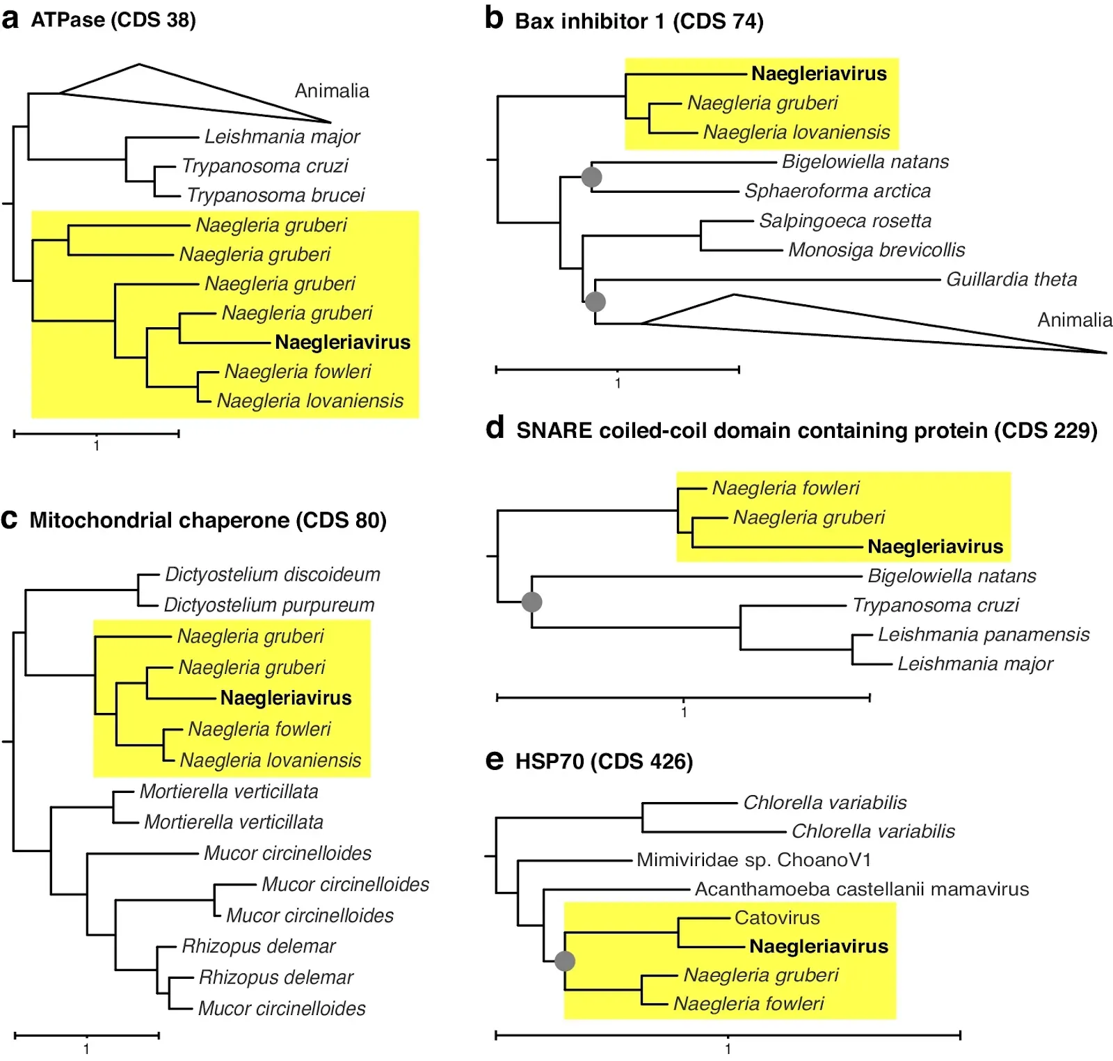 Approximated maximum-likelihood phylogenetic trees are shown for genes encoding an a ATPase-domain containing protein, b Bax 1 inhibition factor 1, c mitochondrial chaperone, d SNARE coiled-coil domain-containing protein, and e HSP70. Bootstrap values lower than 0.8 are depicted as gray circles. Only the relevant subtrees, including Naegleriavirus genes are shown; full versions of the trees are available as Supplementary Files.
Fig. 4: The Naegleriavirus replication cycle in Naegleria clarki. The infection was performed at a multiplicity of infection (MOI) of 10. Fluorescence in situ hybridization (FISH) images are shown. The host cell is depicted in magenta (oligonucleotide probe Nag1088; Supplementary Table 3), with nucleic acid staining by DAPI in cyan. a An uninfected N. clarki trophozoite. b An ameba cell 1 hour post viral infection; small DAPI-stained structures start to accumulate in the cytoplasm. c 4 hpi, intermediate stages of the viral factory are visible. d 8 hpi, the major viral factory and mature viral particles accumulating in the host cytoplasm can be seen. N = nucleus. VF = viral factory. V = virion. Scale bar = 5 µm.
Fig. 5: The ultrastructure of Naegleriavirus virions. Transmission electron microscopy of cryoimmobilized samples processed by freeze substitution or chemically fixed at room temperature (indicated by *). a Medial section of a virion in the host cytoplasm exhibiting the viral core surrounded by membranes, the capsid shell, and a fiber shell. The NiV particle structure is notably similar to that of mimiviruses31. b Medial section of virions showing open and closed stargates (arrowheads). c Virion within a host cell phagosome devoid of its viral core, and membranous structures. Note a well-visible fiber layer covering the electron-dense capsid shell of the virion. d Vertex with starfish-shaped edges (arrowheads). e A lateral section showing a triangular profile of the icosahedral capsid shell surrounded by the fiber layer. All scale bars: 500 nm. CS = capsid shell. CW = core wall. AM = proposed additional membrane. FL = fiber layer. ICS = inner capsid shell. IM = inner membrane. VC = viral core. SGC = stargate closed. SGO = stargate opened. Terminology is based on29,30.
Fig. 6: Features of the Naegleriavirus replication cycle.Transmission electron microscopy of cryoimmobilized samples processed by freeze substitution or chemically fixed at room temperature (indicated by *). Numbers indicate hours post-infection (hpi). a N. clarki trophozoite forming a phagocytic cup and containing two virions in the phagosome (boxed rectangle); potentially phagocytosed virion marked with an arrowhead. b Open stargate (black arrow head) in the upper virion. Virion underneath displays the beginning of the stargate opening; the extra membrane sac can be seen. c Fusion of a NiV inner membrane with a phagosome membrane (black arrow head). d Two virions devoid of viral cores enclosed by a multilamellar structure within a phagosome. e Fully grown and productive viral factory. f Infected ameba showing a large viral factory and virions accumulating in the host cytoplasm. Membrane-enclosed virions suggest ongoing phagocytosis (white arrowheads). ELS = extracellular lamellar structures. EMS = extra membrane sac. MF = membrane fusion. MLS = multilamellar structure. N = nucleus. NL = nucleolus. PC = phagocytic cup. SG = star gate. VC = viral core. VF = viral factory.
Arthofer, P., Panhölzl, F., Delafont, V. et al.
A giant virus infecting the amoeboflagellate Naegleria.
Nat Commun 15, 3307 (2024). https://doi.org/10.1038/s41467-024-47308-2
Copyright: © 2024 The authors.
Published by Springer Nature Ltd. Open access.
Reprinted under a Creative Commons Attribution 4.0 International license (CC BY 4.0)


The Unintelligent Designer: Refuting The Intelligent Design Hoax

ID is not a problem for science; rather science is a problem for ID. This book shows why. It exposes the fallacy of Intelligent Design by showing that, when examined in detail, biological systems are anything but intelligently designed. They show no signs of a plan and are quite ludicrously complex for whatever can be described as a purpose. The Intelligent Design movement relies on almost total ignorance of biological science and seemingly limitless credulity in its target marks. Its only real appeal appears to be to those who find science too difficult or too much trouble to learn yet want their opinions to be regarded as at least as important as those of scientists and experts in their fields.
Available in Hardcover, Paperback or ebook for Kindle
The Malevolent Designer: Why Nature's God is Not Good

This book presents the reader with multiple examples of why, even if we accept Creationism's putative intelligent designer, any such entity can only be regarded as malevolent, designing ever-more ingenious ways to make life difficult for living things, including humans, for no other reason than the sheer pleasure of doing so. This putative creator has also given other creatures much better things like immune systems, eyesight and ability to regenerate limbs that it could have given to all its creation, including humans, but chose not to. This book will leave creationists with the dilemma of explaining why evolution by natural selection is the only plausible explanation for so many nasty little parasites that doesn't leave their creator looking like an ingenious, sadistic, misanthropic, malevolence finding ever more ways to increase pain and suffering in the world, and not the omnibenevolent, maximally good god that Creationists of all Abrahamic religions believe created everything. As with a previous book by this author, "The Unintelligent Designer: Refuting the Intelligent Design Hoax", this book comprehensively refutes any notion of intelligent design by anything resembling a loving, intelligent and maximally good god. Such evil could not exist in a universe created by such a god. Evil exists, therefore a maximally good, all-knowing, all-loving god does not.
Illustrated by Catherine Webber-Hounslow.
Available in Hardcover, Paperback or ebook for Kindle
Illustrated by Catherine Webber-Hounslow.

No comments :
Post a Comment
Obscene, threatening or obnoxious messages, preaching, abuse and spam will be removed, as will anything by known Internet trolls and stalkers, by known sock-puppet accounts and anything not connected with the post,
A claim made without evidence can be dismissed without evidence. Remember: your opinion is not an established fact unless corroborated.