

A composite image of the Asgard archaeon (inset) found within the microbial mats of Shark Bay, Western Australia.
Image: Iain Duggin, Debnath Ghosal, Brendan Burns
The origin of the eukaryotic cell was one of the most consequential transitions in the history of life. Every animal, plant, fungus and protist is composed of eukaryotic cells—cells containing a nucleus and an elaborate internal organisation that is absent from bacteria and archaea. Without that ancient evolutionary innovation, there would have been no plants, no animals and, of course, no humans.
Yet eukaryotic cells appear not to have arisen through the sudden creation of an entirely new kind of organism. The evidence increasingly indicates that they emerged through an extended evolutionary association between previously independent microorganisms. An archaeal host formed a close relationship with at least one bacterium, and a descendant of an alphaproteobacterium eventually became the mitochondrion—the energy-processing organelle still present in almost every eukaryotic cell. Other cellular features evolved before, during or after this extraordinary merger.
Precisely how that relationship began remains one of evolutionary biology’s most difficult questions. The event occurred billions of years ago, microorganisms rarely leave informative fossils, and the organisms involved have long since disappeared. Scientists must therefore reconstruct the process by combining comparative genomics, cell biology, phylogenetics and observations of living microorganisms whose biology may preserve clues to those ancient interactions.
Now an international research team led by scientists at the University of New South Wales, the University of Technology Sydney and the University of Melbourne has observed something tantalisingly suggestive. As described in an open-access paper in Current Biology, the researchers cultivated a previously unknown member of the Asgard archaea from a microbial mat associated with the stromatolites of Gathaagudu, or Shark Bay, Western Australia.

Using electron cryotomography, the researchers also obtained three-dimensional images of the cells at extraordinary resolution. They found that the archaeon produced chains of membrane-bound vesicles, extracellular fibres and complex internal tube- and cage-like structures. Most strikingly, the archaeal and bacterial cells were seen in direct physical contact through fine intercellular tubes assembled by the bacterium. The two organisms were not merely living in the same microbial community; they were forming an intimate physical association of the kind from which a mutually dependent partnership could evolve. The UNSW account of the discovery describes such microbial mats as a possible “microbial village” in which the ancestors of eukaryotes acquired their characteristic complexity.
This should not be mistaken for scientists having watched the first eukaryotic cell evolve. These are modern organisms, neither species is claimed to be the direct ancestor of eukaryotes, and the bacterial partner is not the alphaproteobacterial lineage that gave rise to mitochondria. Nor has the study established that the observed tubes necessarily transport the metabolites predicted from the genomes. What the researchers have discovered is a plausible living analogue of an early stage in eukaryogenesis: an Asgard archaeon and a bacterium forming the sort of close physical and metabolic association from which more integrated symbiosis could develop. As Brendan Burns and Kymberley Oakley explain in their article in The Conversation, it offers a rare glimpse of what microbial “first contact” might have looked like.
For creationists, this presents several familiar difficulties. The research concerns a natural evolutionary process unfolding among microorganisms on a planet already billions of years old, not the instantaneous creation of plants and animals during a single supernatural creation week. It also shows that the supposedly irreducible complexity of the eukaryotic cell may have been assembled gradually from structures, genes and metabolic capabilities that already existed in simpler organisms.
Most awkwardly of all, the cells composing the human body bear the evidence of that mixed ancestry. Their fundamental architecture is inherited from archaea, while their mitochondria are descendants of bacteria. We are not a separately created “kind” standing apart from the rest of life, but one distant branch of a eukaryotic lineage whose origins lie in intimate partnerships between ancient microbes. The gaps in our knowledge remain real, but—as this research demonstrates—they are questions for investigation, not evidence of magic. Each new discovery makes that gap a little smaller and leaves creationism with correspondingly less darkness in which to conceal its creator.
Brendan Burns' and Kymberley Oakley's article in The Conversation is reproduced here under a Creative Commons licence, reformatted for stylistic consistency:

‘First contact’ that may have led to complex life on Earth finally witnessed by scientists
Microscopic image showing newly discovered Asgard archaeon (Nerearchaeum marumarumayae) derived from microbial mats that offers clues to the formation of complex life.
Debnath Ghosal
On the shores of the west coast of Australia lies a window to our past: the stromatolites and microbial mats of Gathaagudu (Shark Bay).
To the untrained eye they look like a collection of rocks and slime – but they are in fact teeming with microbial life. And these stromatolites are living “relics” of ancient ecosystems that thrived on Earth billions of years ago.
If you wade past, it feels like you’re walking back through time. In fact, the first bubbles of oxygen that filled the atmosphere on early Earth likely came from ancient stromatolites. You could say we owe our very existence to these piles of rocks.
So, what other secrets of our past could these ecosystems tell us? Through decades of research, we know how early life has woven its path through these “living rocks”. But most recently our team embarked on the greatest genealogy search of them all: searching for our great microbial ancestors, the Asgard archaea.
And in a new paper, published today in the journal Current Biology, we report how this search led to the discovery of a key clue that could help explain how complex life evolved on Earth.
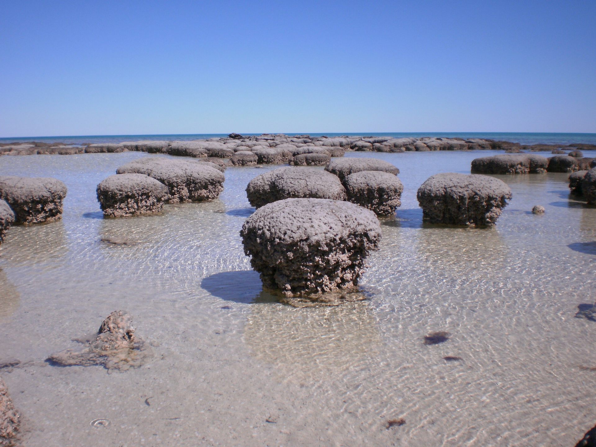
A field of stromatolites in Shark Bay, Western Australia.
Brendan Burns
Asgard archaea were originally named after Norse gods. This fascinating group of microbes sits on the cusp of one of the most significant events in the evolution of life: the origin of the complex cells that make up plants and animals, known as eukaryotes.
Evidence suggests Asgard archaea are the closest relatives of eukaryotes. And that on an early Earth it was the “marriage” of an ancient Asgard archaeon and a bacterium that led to the first eukaryotes.
They formed an ancient partnership. They shared resources and physically interacted, leading to the first complex cells. Like a Romeo and Juliet tale of two distant families coming together, Asgard archaea and bacteria decided it was time to break from traditional family values.
But we have never seen a model of how this may have occurred. Until now.
Holding up a mirror to the ancient past
Our team used the mats of Shark Bay as a “seed” to establish cultures of these ancient microbes. We are one of only four groups worldwide to achieve this, through years of research with a dedicated team of graduate students nurturing the Asgards like offspring.
But the Asgards were not alone. We found them together with a sulphate-loving bacterium. Could this be a model of how complex life may have started on a primitive Earth?
We began by sequencing the Asgards’ DNA to decipher exactly how these microbes tick at the genetic level. We also used artificial intelligence to model how proteins could have behaved in a world before eukaryotes. Evidence suggested these two microbes were sharing nutrients. In other words, they were cooperating.
But we wanted to delve deeper. What do our great microbial ancestors look like? Here we turned to electron cryotomography, a high-resolution imaging approach that allowed us to observe cells and structures at a nanometre scale.
And here we showed – for the first time – an Asgard archaeon and a bacterium directly interacting. Tiny nanotubes were connecting the two organisms – perhaps reflecting what their great-ancestors did on an early Earth that ultimately led to the explosion of complex life as we know it.
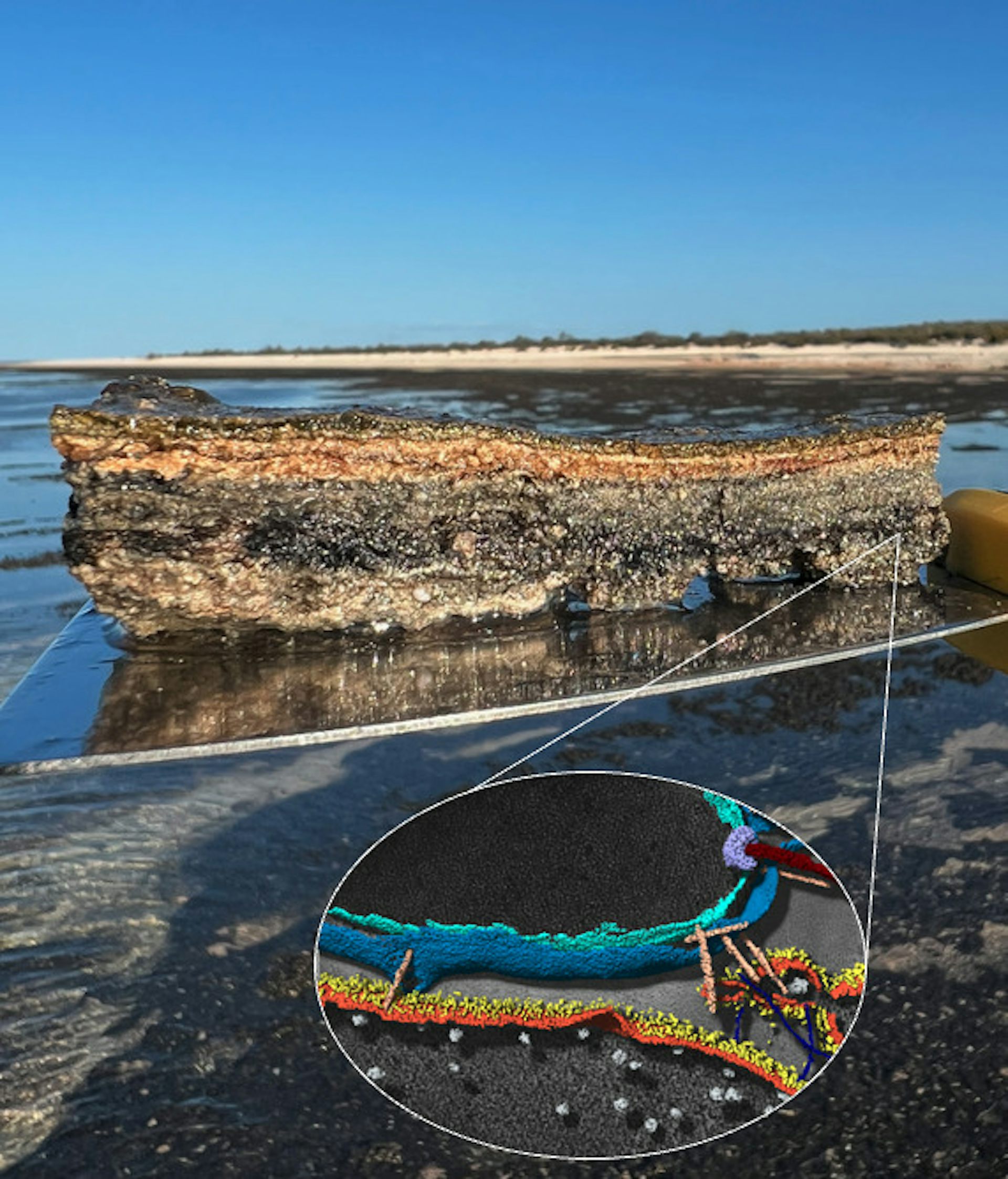
Microbial mat from Gathaagudu (Shark Bay, Australia). Inset: Microscopic image showing Asgard archaeon and bacterium derived from these mats interacting as a model for evolution of complex cells.
Iain Duggin/Bindusmita Paul/Debnath Ghosal/Matthew Johnson/Brendan Burns.
This was a major discovery – one that originated in Gathaagudu, a World Heritage Site with significant environmental and cultural values.
Aboriginal people first inhabited Gathaagudu over 30,000 years ago. We wanted to recognise and celebrate the language of the Malgana people, one of the traditional language groups of Gathaagudu. We also wanted to connect western science with Indigenous Knowledge in a meaningful way.
To this end and working closely with the world’s foremost Malgana language expert, Kymberley Oakley, and Aboriginal elders, a name was granted for our novel Asgard archaeon from the language of the Malgana people: Nerearchaeum marumarumayae. The species name – marumarumayae – is derived from the Aboriginal language of the Malgana people, meaning “ancient home”, a reference to stromatolites being of ancient origin in Earth’s history.
Weaving Aboriginal language into the naming of our new microbe represents a fitting connection between unique Aboriginal culture in Australia and the ancient microbe discovered that calls the mats of Gathaagudu “home”.
Gathaagudu is under threat from global change, from increased heatwaves, cyclonic events and human activity. And among the values to preserve and conserve are the significant Aboriginal connections as well as the trails of life going back through evolutionary time.
With our study we have peered into our past. And maybe like the Montagues and Capulets of Shakespeare, we see distant families of microbes coming together to bridge the divide and ultimately form the early eukaryotes that eventually led to us: a fragile branch on the evolutionary tree of life.

Brendan Paul Burns, Associate Professor, School of Biotech & Biomolecular Science, UNSW and Kymberley Oakley, Indigenous language expert, Indigenous Knowledge
This article is republished from The Conversation under a Creative Commons license. Read the original article.


What the researchers have observed is not the birth of a eukaryotic cell, nor do they claim that either of these living species participated in the original event. It is something more scientifically useful than such an extravagant claim: a living example of how an Asgard archaeon and a bacterium can form the close physical and potentially metabolic association required by plausible models of eukaryogenesis. It turns an otherwise abstract evolutionary scenario into a process that can be observed, analysed and tested.
The discovery also illustrates how major evolutionary innovations need not appear fully formed. Existing cells, membranes, proteins, metabolic pathways and ecological relationships can be recruited and modified as organisms become increasingly interdependent. What eventually became a single complex cell may have begun as cooperation between separate, much simpler cells, with natural selection favouring associations that improved the survival and reproduction of both partners. No foresight was required, and evolution did not need to be aiming towards plants, animals or humans.
For creationists, the implications are profoundly inconvenient. Eukaryotic cells are not evidence of an irreducibly complex creation appearing suddenly and complete. Their structure preserves evidence of descent from archaeal and bacterial ancestors, including the bacterial origin of mitochondria. Every human cell therefore contains evidence that our lineage emerged from microbial evolution and symbiosis over immense periods—billions of years before the few thousand years permitted by a literal reading of Genesis.
There are still important questions about precisely where, when and in what sequence eukaryotic features evolved. Science acknowledges those uncertainties and investigates them; it does not disguise ignorance as certainty or insert a supernatural agent into whatever remains unexplained. This research has not closed every gap in the story of eukaryogenesis, but it has made one possible stage more tangible and more testable. Once again, progress came from treating complexity as the product of natural history—not as evidence that history was interrupted by magic.
Advertisement
Amazon

Amazon

Amazon

Amazon

Amazon

Amazon

Amazon

Amazon

Amazon

Amazon

Amazon

Amazon

All titles available in paperback, hardcover, ebook for Kindle and audio format.
Prices correct at time of publication. for current prices.



No comments :
Post a Comment
Obscene, threatening or obnoxious messages, preaching, abuse and spam will be removed, as will anything by known Internet trolls and stalkers, by known sock-puppet accounts and anything not connected with the post,
A claim made without evidence can be dismissed without evidence. Remember: your opinion is not an established fact unless corroborated.